
")

The package is a capture, analysis and DICOM image management system dedicated to radiotherapy.
It consists of a series of software modules associated with a radiotherapy PACS, which can provide a unique tool for managing all the data needed for the radiotherapy workflow.The system is able to:
- receive from PACS the diagnostic images of the patients to be treated
- send diagnostic images to radiotherapy workstations
- archive the treatment plans generated by such workstations
- visualize DICOM-RT objects related to treatment plans
- archive and visualize DICOM objects associated with non-DICOM (PDF Embedded) document management
The system has been tested with various TPS treatment plans including RayStation, Pinnacle, Monaco, MIMVista, TomoTherapy, Eclipse, Oncentra e CyberKnife.
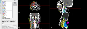
On this platform, a segmentation station has been equipped with all the multimodal volumes visualization and management tools required by the case. Such station allows visualization and management of images obtained with different technologies (CT, PET MRI, 3DUS) and allows the user to manage segmentations performed or uploaded by a database and the use of aberration correction and algorithms segmentation developed during the project.
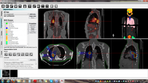
Figure 1: Example of the segmentation user interface developed within the project.
The following are the characteristics of the workstation.
Module for DICOM files managementSCU/SCP query/retrieve, SCU storage, radioterapy SOP Class management, advanced forwanding rules of study / series to other DICOM nodes (with anonymization possibility), loading DICOM files from DICOMDIR and Filesystem, creation of Patient CD on CD/DVD/USB, DICOM objects import/export, execution of algorithms (US correction) on the reception event of the series, DICOM and user configurator server.
Module for viewing and fusion2D and 3D display and multimodal display, 2D/3D views managing, measuring instruments (line, angle, area), TAGs DICOM visualization of loaded series, screenshots printing and salving, structures and doses visualization, manual recording tools (rigid), possibility of connecting to external plugins for multimodal recording
Segmentation module3D native segmentation, 2D and 3D manual segmentation tools, export / import segmentation in DICOM RTSTRUCT format
External segmentation plugins2D and 3D semi-automatic segmentation on PET images, automatic segmentation on co-recorded images US-CT, CT-PET, etc. Algorithm of automatic selection (super partes algorithm) of the segmentation algorithm.
